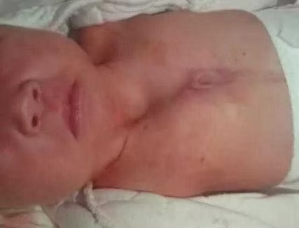
The patient is a 5-month-old female infant born with a longitudinal midline scar extending from the chest to the abdomen, along with a congenital absence of the bony structures of both the upper sternum and the upper abdominal wall. Since birth, she has experienced two episodes of pneumonia, both complicated by severe respiratory distress. Her condition stabilized after emergent medical intervention.
Previous Case
Revision Surgery for Nuss Procedure Fail...
Next Case
Simultaneous Surgery for A One-month-old...
Medical History
Preoperative Examination
There is a prominent longitudinal scar along the midline of both the anterior chest wall and the abdominal wall, extending from the chest wall to the umbilicus. The upper part of the sternum is split along the midline, with only a minimal bony structure remaining connected at the very bottom of the sternum (above the xiphoid process), forming a "V"-shaped defect. Meanwhile, the upper abdominal wall is also split along the midline, resulting in an inverted "V"-shaped defect. Additionally, paradoxical breathing is evident in both the upper chest wall and upper abdominal region. The echocardiography shows no structural cardiac abnormalities. The child was ultimately diagnosed with sternal cleft combined with abdominal wall defect.
Surgical Overview
A midline incision was made along the anterior chest wall, beginning from the sternoclavicular joints and extending downward to the upper abdominal wall. For the sternal defect area, reconstruction was performed using the MatrixRIB bars. The ends of the bars were precisely fixed onto the residual bony structure at the lower end of the sternum. Meanwhile, appropriate reinforcement was carried out at the costal cartilages to enhance stability.
After eliminating the chest wall defect as much as possible, a patch was placed inside the thoracic cavity to prevent adhesion between the bars and thoracic organs. Subsequently, another patch was applied over the surface of the thoracic cavity to further reinforce the bars and ensure the effectiveness of the reconstruction.
For the defect in the upper abdominal wall, the same approach as for the chest wall defect was employed, utilizing two patches for reconstruction and reinforcement. Finally, the edges of the patch covering the surface of the abdominal wall were securely fixed to the costal arch to maintain its stability.
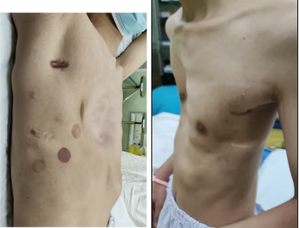
The surgery was completed successfully without complications. Postoperatively, the appearance of the chest and abdominal wall returned to normal, and paradoxical breathing was completely resolved.
Related Photos