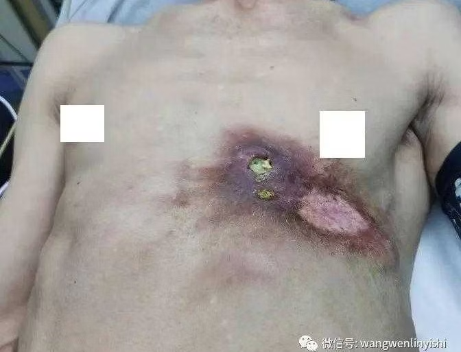
The patient is a 67-year-old man who underwent surgery and radiation therapy for the chest wall tumor 35 years ago. After the radiation therapy, the skin at the treated area darkened but showed no ulceration. Regular follow-ups over the years confirmed no recurrence of the tumor. However, two months ago, the patient suddenly observed redness, swelling, and ulceration at the surgical site, accompanied by discharge oozing out.

Previous Case
Treatment for A 2-Month-Old Infant with ...
Next Case
Repair and Reconstruction of Chest Wall ...
Medical History
Preoperative Examination
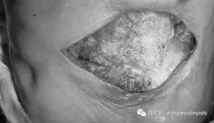
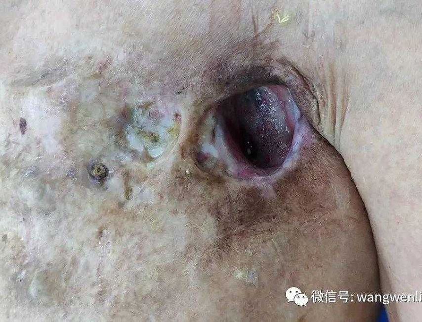
The left anterior chest wall shows a large area of chronic radiation damage, featuring fistulas accompanied by some grayish-white discharge. The overlying skin is dark brown in color. Deep within the fistulas, an obvious necrotic lesion is present, with adhesions to the pericardium. The patient was diagnosed with chest wall defect resulting from radiation therapy for the chest wall tumor.
Surgical Overview
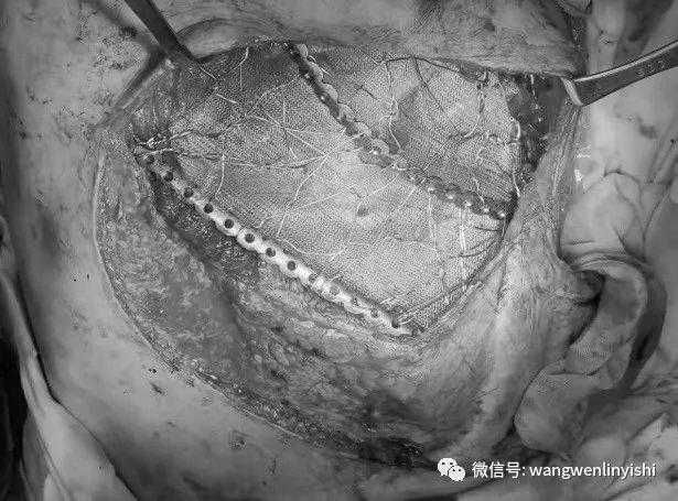
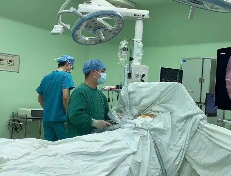
Learn about the surgical case of a 67-year-old man with a chest wall defect resulting from previous tumor surgery and radiation therapy. After developing redness, swelling, and ulceration at the surgical site, he was diagnosed with chest wall infection. The surgery involved excising necrotic tissue, reconstructing the chest wall with MatrixRIBs, and using pedicled flaps for coverage. Successful outcomes were achieved, restoring the integrity of the chest wall.
Related Photos